Most people picture an eye exam as reading letters off a chart until the print gets too small to make out. That single test is only a small part of what actually happens once a patient sits down in the exam chair.
A comprehensive eye exam looks at far more than sharpness of vision. Keep reading to learn what a comprehensive eye exam checks for and why each part of the process matters for long-term vision.
Why an Eye Exam Involves More Than Reading a Chart
A typical visit starts with a technician who takes your history, checks visual acuity, and runs preliminary tests before your eye doctor enters the room. From there, your eye doctor examines the structures inside and around the eye, often after dilating the pupils for a wider view. Each step is designed to catch a different type of problem, since some conditions affect focus while others develop with no early symptoms at all.
What Your Eye Doctor Evaluates During a Comprehensive Exam
The list below covers the core areas most eye doctors assess during a routine visit.
1. Visual Acuity and Refraction
The eye chart test measures how clearly a patient sees at a distance, but the subsequent refraction determines the exact lens power needed to correct vision. Prescriptions shift gradually over time, and this part of the exam is what confirms whether current glasses or contacts still match a patient’s eyes.
2. Eye Pressure and Signs of Glaucoma
A quick pressure check, called tonometry, measures the intraocular pressure. Elevated pressure can be an early indicator of glaucoma, a condition that damages the optic nerve so gradually that many people lose peripheral vision before noticing anything is wrong. Catching that pressure change early is often the only way to intervene before permanent damage occurs.
3. The Lens and Early Cataract Changes
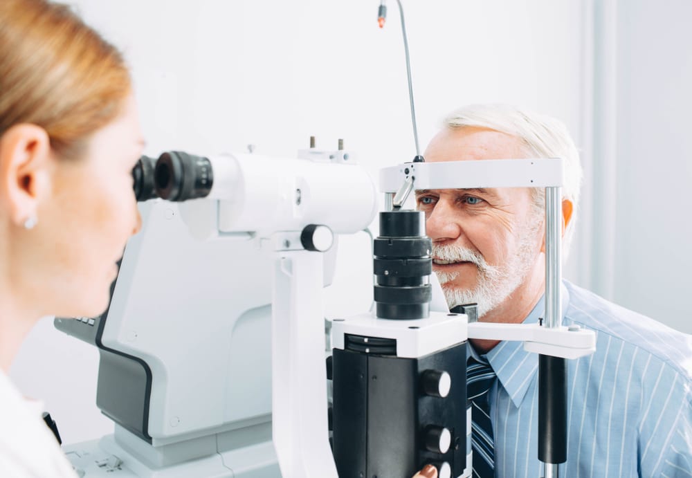
With a special microscope called a slit-lamp, your eye doctor inspects the eye’s natural lens for any signs of cloudiness. Early cataracts often go unnoticed, but this careful check can reveal them long before you notice blurry or hazy vision.
4. The Retina and Macula
Once the pupils are dilated, the doctor gets a clear view of the retina, optic nerve, and blood vessels at the back of the eye. This is where early signs of diabetic changes or macular degeneration often appear, well before a patient notices any change in central vision.
5. Overall Eye Health and Surface Conditions
The exam also covers the eyelids, tear film, and surface of the eye. Dry eye, styes, and other surface conditions are common findings, and while they are rarely serious, they can affect comfort and contact lens wear if left unaddressed.
When to See an Eye Doctor About Your Next Exam
An annual exam is generally recommended for most adults, though anyone with diabetes, a family history of eye disease, or existing vision correction may benefit from more frequent visits. The experienced ophthalmologists at Colorado Eye Consultants combine a full range of diagnostic testing with a straightforward explanation of what each result means for a patient’s vision.
Due for your next comprehensive eye exam? Schedule an appointment at Colorado Eye Consultants in Littleton, CO, today.